
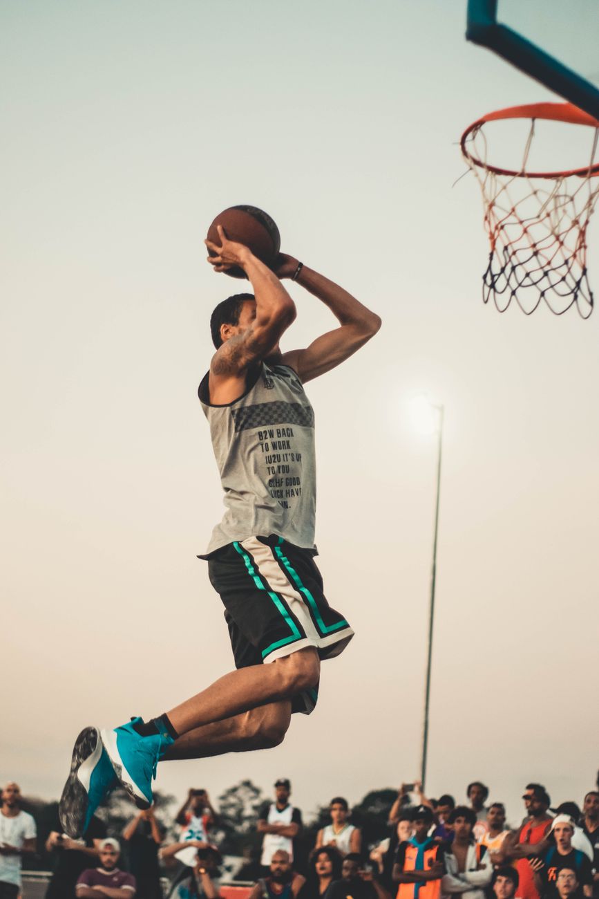
Patellar Instability care in North Texas. Dr. Manoj P. Reddy evaluates and treats this condition at his Dallas, Rockwall and Garland offices.
What is patellar instability?
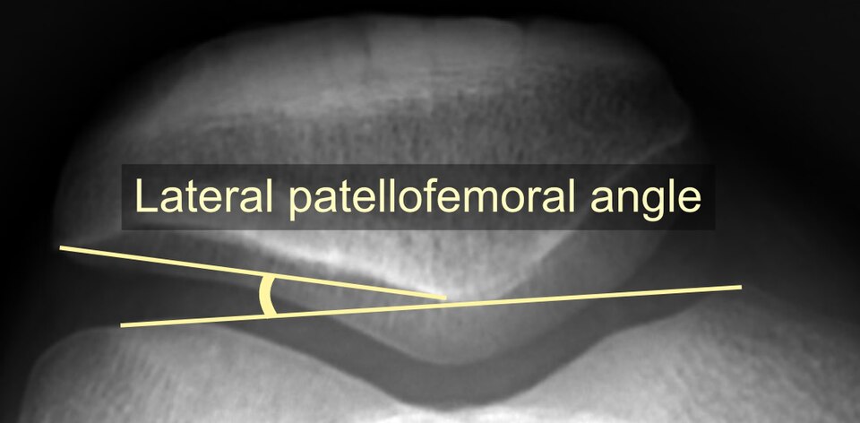
The kneecap (patella) glides in a groove at the front of the thigh bone. Patellar instability means the kneecap slips partly (subluxation) or completely (dislocation) out of that groove, almost always toward the outside of the knee.
What causes it?
A first dislocation usually happens with a twist on a planted foot or a direct blow. It typically tears the medial patellofemoral ligament (MPFL), the main restraint on the inner side. Some people are more prone because of their anatomy:
- A shallow groove (trochlear dysplasia)
- A high-riding kneecap
- Knock-kneed alignment or rotation of the leg bones
- Generalized loose ligaments
What are the symptoms?
- A feeling of the kneecap shifting or the knee giving way
- Pain at the front of the knee
- Swelling after an episode
- A visible deformity if the kneecap stays out of place
How is it diagnosed?
By examination, X-rays and an MRI. The MRI shows the torn ligament and any cartilage fragment knocked loose. Measurements of the groove, kneecap height and alignment help predict the risk of recurrence.
How is patellar instability treated?
A first dislocation without a loose fragment is usually treated without surgery: a short period in a brace, then physical therapy to strengthen the quadriceps and hip muscles.
Surgery is recommended for recurrent dislocations, or after a first dislocation when a piece of cartilage and bone has broken off. MPFL reconstruction rebuilds the torn ligament with a tendon graft. When alignment contributes, a tibial tubercle osteotomy shifts the patellar tendon attachment to improve tracking.
What is recovery like?
After MPFL reconstruction, a brace and crutches are used early and motion is restored over the first weeks. Return to sport is typically around four to six months, and longer when an osteotomy is added.
This page is general information, not medical advice. Recovery times are typical ranges and vary from person to person.
KNEE CONDITIONS
All knee topics
ACL Tears
Meniscus Tears
Cartilage Injuries
Multi-Ligament Injuries
Patellar Instability
Quadriceps and Patellar Tendon Tears
Arthritis
Malalignment
AT A GLANCE
Manoj P. Reddy, MD
- Board-certified orthopaedic surgeon, fellowship-trained in sports medicine and shoulder surgery at the Rothman Orthopaedic Institute
- Orthopaedic surgery residency at the University of Chicago
- Specializes in arthroscopic, reconstructive and joint replacement surgery of the shoulder, knee and elbow
- Assistant team physician for the Philadelphia 76ers, Eagles and Phillies during fellowship; covers Rowlett, Garland and Naaman Forest high schools
- Author of 27 peer-reviewed publications in sports medicine
- Living Magazine Top Doctor, 2024

