
MPFL Reconstruction in North Texas. Dr. Manoj P. Reddy offers this treatment at his Dallas, Rockwall and Garland offices.
What is the MPFL?
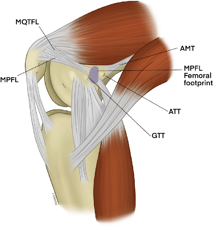
The medial patellofemoral ligament (MPFL) is a band of tissue that runs from the inner edge of the kneecap to the inner side of the thigh bone. It is the main restraint that stops the kneecap from sliding toward the outside of the knee, particularly in the first 30 degrees of bending, before the kneecap is fully seated in its groove.
How is the MPFL injured?
The MPFL tears or stretches in almost every kneecap dislocation. Once it has healed in a lengthened position it no longer holds the kneecap securely, which is why a first dislocation is often followed by others.
Who is a candidate for MPFL reconstruction?
- Two or more kneecap dislocations
- Ongoing instability or apprehension despite a full course of physical therapy
- A first dislocation with a loose fragment of cartilage and bone that needs to be fixed or removed
- Younger patients with anatomic risk factors that make another dislocation likely
Before surgery, X-rays and an MRI are used to measure the shape of the groove, the height of the kneecap and the alignment of the patellar tendon. If these are significantly abnormal, a tibial tubercle osteotomy may be added so that the new ligament is not overloaded.
How is the surgery performed?
The knee is first examined arthroscopically and any cartilage damage is treated. A tendon graft, either one of your own hamstring tendons or a donor tendon, is then attached to the upper inner edge of the kneecap and passed beneath the skin to the thigh bone. It is fixed at the precise point where the original ligament attached, confirmed with X-ray during the operation, and tensioned so the kneecap is guided into the groove without being pulled too tight.
What is recovery like?
Surgery is outpatient. A brace is worn for walking for about six weeks, initially locked straight, and crutches are used until the thigh muscle is working well. Bending is increased steadily over the first weeks. Strengthening of the quadriceps and hip muscles is the focus of therapy.
Jogging usually begins at around three to four months, and return to cutting and pivoting sports is typically at four to six months, after strength testing.
Download the MPFL reconstruction protocol.
How successful is it?
MPFL reconstruction reliably prevents further dislocations in the large majority of patients, and most return to sport. Results are best when contributing alignment problems are identified and addressed at the same time.
What are the risks?
Specific risks include stiffness, pain or pressure at the front of the knee if the graft is too tight, fracture of the kneecap at the fixation site, and another dislocation. As with any operation, there are also general risks of infection, blood clots, stiffness, nerve or blood vessel injury and problems with anesthesia. These are uncommon, and steps are taken to lower them.
This page is general information, not medical advice. Recovery times are typical ranges and vary from person to person.
KNEE TREATMENTS
ACL and Ligament Surgery
ACL Reconstruction and Revision
PCL Reconstruction
MCL Reconstruction
Multi-Ligament Reconstruction
Meniscus Surgery
Meniscus Repair and Meniscectomy
Meniscus Root Repair
Meniscus Transplant
Cartilage and Realignment
Cartilage Restoration
MACI Cartilage Implantation
Osteochondral Allograft (OCA)
Knee Osteotomy
Kneecap Stabilization
Patellar Stabilization
MPFL Reconstruction
Tibial Tubercle Osteotomy
Other Knee Treatments
Knee Arthroscopy
Quadriceps and Patellar Tendon Repair
Partial Knee Replacement
Injections
AT A GLANCE
Manoj P. Reddy, MD
- Board-certified orthopaedic surgeon, fellowship-trained in sports medicine and shoulder surgery at the Rothman Orthopaedic Institute
- Orthopaedic surgery residency at the University of Chicago
- Specializes in arthroscopic, reconstructive and joint replacement surgery of the shoulder, knee and elbow
- Assistant team physician for the Philadelphia 76ers, Eagles and Phillies during fellowship; covers Rowlett, Garland and Naaman Forest high schools
- Author of 27 peer-reviewed publications in sports medicine
- Living Magazine Top Doctor, 2024

